
Laboratory for Engineering of Early Cancer Diagnosis
- Laboratory type:
- teaching and research-expert laboratory
- Description of laboratory activities:
- characterization and early diagnosis, as well as screening of biological tissues using OMIS devices, FTIR, and UV/Vis/NIR spectroscopy
The Laboratory for Engineering of Early Cancer Diagnosis has been successfully developing and improving cancer detection methods for many years. The Department of Biomedical Engineering developed and patented the Optomagnetic Imaging Spectroscopy (OMIS) method, authored by Dr. Đuro Koruga, retired Full Professor, and Dr. Aleksandar Tomić, Scientific Associate, which provides significant contributions to the characterization of materials, with an emphasis on biological materials. Today, this method, in its improved form, with new hardware and software solutions, achieves significant and high accuracy rates in detecting various types of cancer.
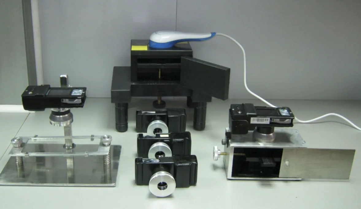
One of the significant contributions of the Department of Biomedical Engineering is the development of a method and device construction for early diagnosis of cervical cancer, which serves for quick, simple, and painless detection of abnormal cells in samples taken by swabs from the cervix and cervical canal. This device serves as a screening and diagnostic method, and due to its simplicity, it can be used in health centers to relieve specialized hospitals and screen a large number of women. The device was developed as an improved version of the original OMIS device as part of the project by the Ministry of Education, Science, and Technological Development of the Republic of Serbia III 41006 titled "Development of new methods and techniques for early diagnosis of cervical, colorectal, oral cavity, and melanoma cancer based on digital images and excitation-emission spectra in the visible and infrared domains."
In addition to examining cervical cells to detect cancer at an early stage, the Laboratory for Engineering of Early Cancer Diagnosis also examined tissue samples of the colon and oral cavity in vitro and skin in vivo, as well as other biological tissues. From the mentioned research conducted within the project, in collaboration with the Faculty of Medicine, University of Belgrade, the Faculty of Dentistry, University of Belgrade, and GAK Narodni Front, the following doctoral dissertations emerged:
- D. Kojić (2010): “Characterization of structural, mechanical, electrical, and magnetic properties of skin using optical and nanoprobe microscopes“,
- B. Jeftić (2017): "Algorithm for determining the biophysical state of epithelial tissue based on spectroscopy",
- A. Dragičević (2018): "Biomechanical and optical characterization of epidermal tissue",
- B. Lisul (2019): "Application of optomagnetic imaging spectroscopy in biophysical characterization of marginal and cancer-altered tissue in the oral cavity",
These contributed the following results in early cancer detection:
scale 70–100%| Tissue | Position on the scale | Result |
|---|---|---|
| Skin – in vivo: | 90-92% | |
| Cervix – in vitro (PAP II – PAP V slides): | 94-96% | |
| Colon tissues – ex vivo: | 92-96% | |
| Oral cavity – in vitro: | 82-84% | |
708090100 |